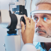
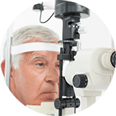
Micropulse Laser Therapy
Micropulse laser therapy is offered for suitable cases of diabetic macular edema, central serous retinopathy (CSR), and early glaucoma. Instead of delivering continuous energy, the laser emits short pulses with rest intervals, allowing tissue to cool between applications. This approach reduces the risk of collateral damage while still achieving the desired therapeutic effect. At present, International Eye Hospital provides the only Micropulse system of its kind in Pakistan.
YAG Laser Treatment
YAG laser is used to treat posterior capsular opacification (secondary cataract) and selected forms of angle-closure glaucoma. In posterior capsulotomy, a focused laser opening is created behind the intraocular lens to restore visual clarity after cataract surgery. In iridotomy, the laser creates a small passage in the iris to improve fluid outflow and reduce pressure in narrow-angle eyes. Both procedures are non-incisional and usually completed within a short outpatient visit.
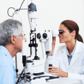
Argon Laser Therapy
Argon laser therapy is a key tool in the management of diabetic retinopathy, retinal tears, and certain vein occlusions. The laser is used to seal leaking vessels, reinforce weak areas, and secure the retina where there is a risk of detachment. Treatment is planned according to the location and extent of disease, and may be delivered in focal or panretinal patterns. The objective is to stabilise the retina and lower the chance of further vision-threatening events.
Yellow Laser (577 nm)
Yellow (577 nm) laser is particularly suited for work in and around the macula. Its wavelength allows targeted treatment of focal leaks, diabetic macular edema, and specific vascular abnormalities while limiting heat spread to adjacent tissue. This makes it a preferred option for selected cases where precision near the centre of vision is critical. Treatment parameters are tailored to the patient’s condition, imaging findings, and response over time.
Biometry – IOL
Master 700
For cataract surgery planning, we use the IOL Master 700, widely regarded as a gold-standard biometry system. It provides highly accurate measurements of the eye, allowing precise selection of intraocular lens power. This contributes to sharp, customised visual outcomes after cataract surgery.

Fundus Photography &
Retinal Imaging
Fundus photography and retinal imaging capture high-resolution images of the back of the eye. These are used to document and monitor diabetic changes, macular disease, retinal tears, and other retinal conditions. The images are essential for accurate diagnosis, treatment planning, and long-term follow-up.

Please call or book an appointment to discuss laser treatment.
