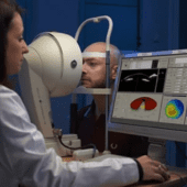
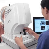
OCT Scan (Optical Coherence Tomography)
Our OCT system uses Swept-Source Technology, scanning at 100,000 scans per second to capture clear, detailed images of the deepest layers of the eye. With its 1050 nm wavelength, it can visualise the retina, vitreous, choroid, and even the sclera with exceptional clarity. It also provides true-colour fundus imaging and wide-field views. The scan is quick, painless, non-contact, and offers critical information for diagnosing macular and retinal disorders with high precision.
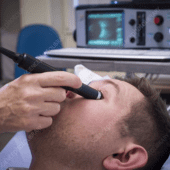
B-Scan
Ultrasound
B-scan ultrasound provides detailed internal imaging when the retina cannot be seen directly, such as in cataract, vitreous haemorrhage, or corneal opacity. It is essential for diagnosing retinal detachments, intraocular tumours, vitreous haemorrhage, and other posterior segment disorders. The test is quick, safe, and a valuable part of assessment in complex cases.
Visual
Field Testing
Visual field testing measures peripheral vision to detect early glaucoma, optic nerve damage, and certain neurological conditions. It generates detailed maps that help in diagnosing disease and monitoring progression over time. Regular visual field analysis is an important component in the long-term management of glaucoma and related disorders.
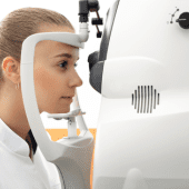
Corneal Topography &
Tomography
Corneal topography & tomography analyse the shape, thickness, & curvature of the cornea. These tests are essential for screening patients for SMILE Pro, Femto LASIK, & Presbyond, and for diagnosing keratoconus. They provide precise three-dimensional mapping within seconds, supporting safe planning for refractive & other corneal procedures.
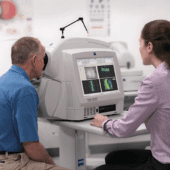
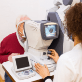
Biometry – IOL
Master 700
For cataract surgery planning, we use the IOL Master 700, widely regarded as a gold-standard biometry system. It provides highly accurate measurements of the eye, allowing precise selection of intraocular lens power. This contributes to sharp, customised visual outcomes after cataract surgery.
Fundus Photography &
Retinal Imaging
Fundus photography and retinal imaging capture high-resolution images of the back of the eye. These are used to document and monitor diabetic changes, macular disease, retinal tears, and other retinal conditions. The images are essential for accurate diagnosis, treatment planning, and long-term follow-up.
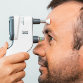
Eye Pressure
Testing
Tonometry measures intraocular pressure, a key parameter in the diagnosis and management of glaucoma. Both air-puff and applanation methods are used to obtain highly accurate and consistent readings. Regular pressure checks help in assessing risk, and monitoring response effectively over time.
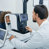
Auto-Refractor &
Keratometer
The auto-refractor and keratometer provide quick, accurate measurements of refractive error and corneal curvature. These readings are essential for prescribing glasses and contact lenses, and for planning refractive and cataract surgeries. The process is fast, and comfortable for
patients.

Fundus Photography & Retinal Imaging
Fundus photography and retinal imaging capture high-resolution images of the back of the eye. These are used to document and monitor diabetic changes, macular disease, retinal tears, and other retinal conditions. The images are essential for accurate diagnosis, treatment planning, and long-term follow-up.
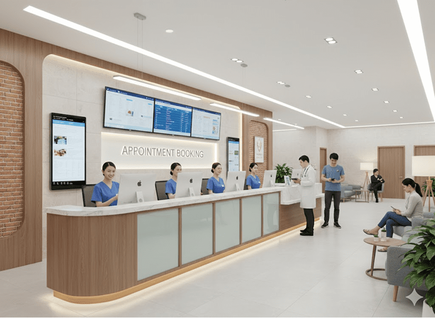
Reach out to our team to book your diagnostic evaluation.
